|
What is Coronary Angiography?The word Angio actually comes from the Greek word Angeio which means blood vessel or vessel. Graphy means the writing of...or representation in some manner...such as picture or audio format of a specific object...in this case, Coronary Arteries.
Heart Catheterization - the advancement and positioning of a flexible tube into one of the hearts chambers was performed by a Dr Werner Forssmann...a German physician...on himself!...using a urinary catheter. The year was 1929. Prior to that date there had been some experimentation but unlike other efforts Dr Forssman actually used X-ray equipment to guide and position the catheter...documenting his efforts with a chest X-ray. He was immediately fired for what seemed at the time like reckless experimentation...he did end up receiving a Nobel prize though in 1956. Some compensation perhaps. Left Heart Catheterization...along with Coronary Angiography did not occur until the late 1950s with experimentation to selectively guide a catheter tip into the origins of the coronary arteries. The realization and potential was seen after an accidental occurrence...a catheter kind of just jumped in there!...as it was in the anatomical neighborhood! A Cardiologist by the name of Mason Sones is credited with discovering and realizing the potential of selectively taking pictures of the Coronary Arteries and establishing a technique for others to follow...and the rest as they say is history! Many technological advancements have been made between then and now, but essentially the procedure for Coronary Angiography remains the same.
In a nutshell Coronary Angiography involves the introduction of a catheter...a small diameter plastic tube...into the arterial system. The catheter is advanced and manipulated under X-ray guidance into the origin(s) of the coronary arteries which open from the Aortic root...just above the Aortic valve (remember from your biology class!) Once a catheter tip is sitting correctly in a coronary artery origin, a few milliliters of X-ray contrast media is pushed through the catheter and into the selected coronary artery. The contrast fills the coronary artery for a few seconds and moving X-ray pictures are acquired...recording a true real-time representation of the coronary artery. Any coronary artery abnormalities such as a reduced diameter due to the build up of calcified plaque which is the process of atherosclerosis...and thrombus which is a blood clot can easily and immediately be assessed from coronary angiogram images.
Three different catheters of the same length and diameter...but with very different catheter tip shapes are utilized in a Coronary Angiogram procedure. The three different shapes are necessary to assist the manipulation of the catheter tip into the correct position. Catheters can only be steered using forwards, backwards, clockwise and counter-clockwise movements...so of course some pre shaping at manufacture is hugely beneficial...along with a slick technique by the attending Cardiologist of course.
Coronary Angiogram sequence.
Does the presence of symptom-less disease mean that a person is well and healthy? ....more on this Click Here.... Click here to go from Coronary Angiography to Coronary Heart Health Home Page
|
| Achieve Your Goal Fat loss Quickly Understand the No Gimmicks!
|
|
| Weight Control Hot Links |
| Make keeping healthy THE priority |


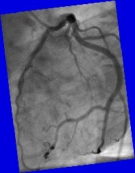
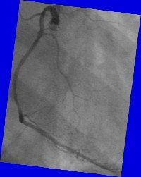
 Here's a still shot of the Right Coronary Artery. This artery usually has one main branch for most of its length terminating in a cluster of three or four branches..not visible in this image. During a Coronary Angiogram, the right coronary artery is the one that looks like a letter C or L whereas the left coronary arteries tend to look a bit like a set of spiders legs!


